Shopping cart

Subtotal 0

View cartCheckout

USG / Ultrasound Jodhpur

See What Others Miss,
Diagnose with Precision

USG and advanced sonography provide safe, real-time imaging of the uterus, ovaries, follicles, pregnancy and pelvic organs.

Our specialists use ultrasound for fertility evaluation, follicle monitoring, pregnancy assessment and diagnosis of conditions like PCOS, fibroids or cysts.

It gives clear, non-invasive answers that support faster and more accurate treatment decisions.

Are You Facing These Issues?

Free Consultation
from our Consultants

When Imaging Becomes the Essential Answer

When Is USG and Advanced
Sonography Recommended?

USG and advanced sonography are recommended across a wide range of clinical situations in women, from investigating the cause
of infertility and recurrent pregnancy loss to monitoring every critical milestone of a healthy pregnancy.

Difficulty Conceiving or Unexplained Infertility

When a woman has been trying to conceive for 6 to 12 months without success and basic blood tests have not identified a clear cause, pelvic ultrasound is the essential next step to evaluate the uterus, ovaries, endometrium, and follicular development.

Suspected PCOS, Endometriosis, or Uterine Fibroids

When a woman has been trying to conceive for 6 to 12 months without success and basic blood tests have not identified a clear cause, pelvic ultrasound is the essential next step to evaluate the uterus, ovaries, endometrium, and follicular development.

Recurrent Miscarriages or Failed IVF Cycles

When a woman has experienced repeated pregnancy loss or multiple failed IVF cycles, 3D uterine sonography is critical for identifying hidden structural causes such as uterine septum, submucous fibroids, polyps, or congenital anomalies that standard 2D scans can overlook.

Fertility Treatment Monitoring

When a woman is undergoing ovulation induction, IUI, or IVF, serial follicle monitoring ultrasounds track the development of ovarian follicles, confirm the trigger injection timing, and assess endometrial receptivity at each critical stage of the treatment cycle.

Pregnancy Confirmation and Viability Assessment

When a pregnancy has been confirmed, an early ultrasound at 6 to 8 weeks confirms viability, detects the fetal heartbeat, identifies the number of embryos, and rules out ectopic pregnancy before the next stage of antenatal care begins.

High-Risk Pregnancy and Third Trimester Monitoring

When a pregnancy is high-risk due to hypertension, gestational diabetes, twin pregnancy, or a history of complications, Color Doppler monitoring of umbilical artery blood flow and serial growth scans assess placental function, fetal wellbeing, and intrauterine growth restriction.

Prepared, Performed, and Precisely Reported

How USG and Advanced Sonography Are Performed

Every USG and sonography scan at Galwa Care Hospital follows a structured, patient-friendly process designed to ensure the most accurate diagnostic images with the greatest possible comfort for every woman.

Preparation for Abdominal USG

Drink 4 to 6 glasses of water one hour before a standard abdominal pelvic scan as a full bladder gives significantly clearer images of the uterus and ovaries. Wear loose, comfortable two-piece clothing for easy access and bring all previous scan reports and referral documents.

Preparation for Transvaginal USG (TVS)

Empty your bladder completely before a transvaginal scan as no full bladder is needed for this technique. The procedure uses a slim, lubricated probe and is safe, quick, and provides a significantly clearer view of the uterus and ovaries than abdominal scanning alone.

Preparation for 3D and 4D Sonography

Best scheduled between 26 and 32 weeks for optimal fetal facial images with adequate amniotic fluid. Stay well-hydrated in the days before for clearer amniotic fluid levels. Have a light meal 1 to 2 hours prior to encourage baby movement during the scan session.

The Scanning Procedure

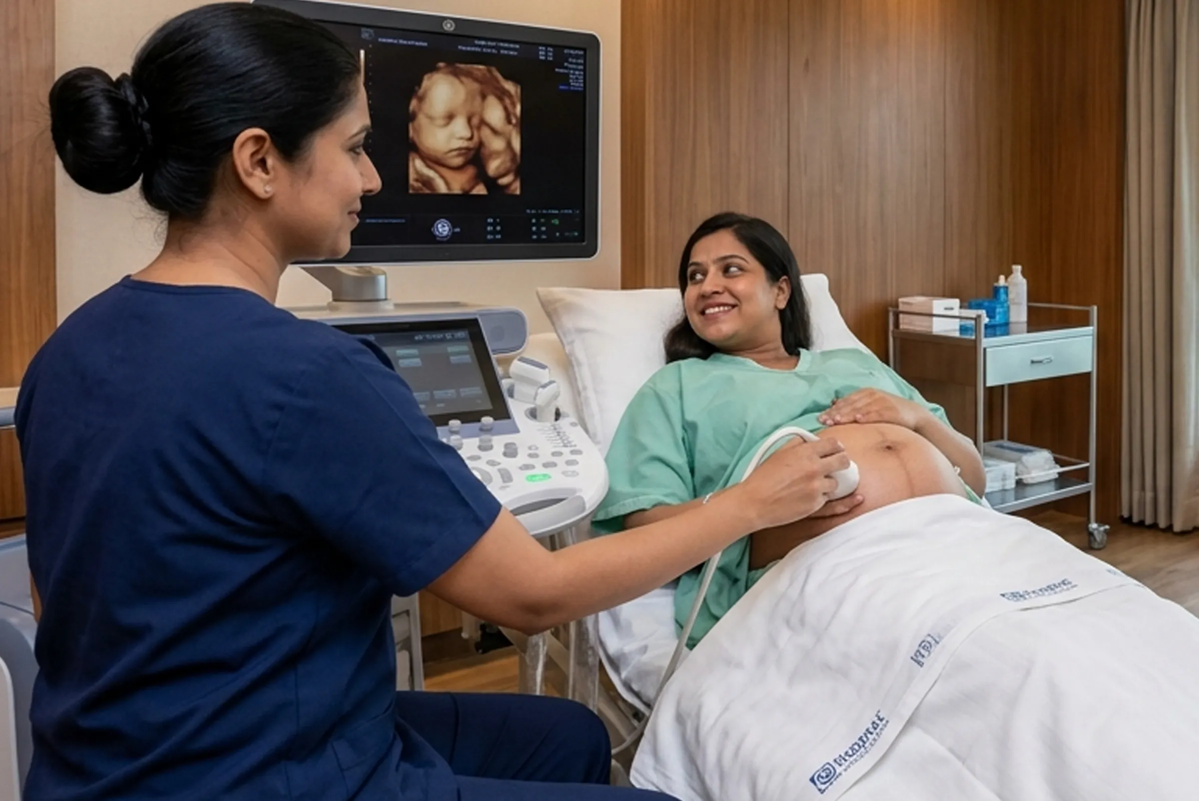
You are positioned comfortably on the examination couch. A clear gel is applied to the abdomen for abdominal scanning, or a slim probe is gently inserted for transvaginal imaging. The sonologist moves the transducer carefully to capture comprehensive images of all relevant structures from every required angle.

3D, 4D, and Color Doppler Imaging

For 3D and 4D scans, multiple simultaneous scan frames are combined to create a three-dimensional image or a real-time live video of the baby. For Color Doppler, blood flow through the uterine arteries, ovarian vessels, umbilical artery, and fetal circulation is mapped in vivid colour-coded real-time imaging.

Clinical Interpretation and Report

Every scan at Galwa Care Hospital is interpreted by an experienced sonologist or radiologist working in coordination with your gynaecologist or fertility specialist. A detailed written report is prepared and explained to you clearly, ensuring the findings translate into actionable clinical guidance.

Same-Day Report and Specialist Consultation

All USG, 3D, 4D, and Color Doppler reports are provided on the same day of the scan. A specialist consultation is available to walk you through your results, answer your questions, and connect your findings directly to your fertility treatment or antenatal care plan.

Clarity That Changes the Course of Care

Benefits of USG and Advanced Sonography

Advanced sonography at Galwa Care Hospital provides the most comprehensive, non-invasive window into female reproductive
health and fetal development, enabling faster diagnosis, more targeted treatment, and better outcomes for every patient.

Completely Safe and Non-Invasive

USG uses high-frequency sound waves, not radiation, making it completely safe for women of all ages, for pregnant women at every trimester, and for the developing baby throughout the entire pregnancy.

Identifies Hidden Causes of Infertility

Pelvic USG and 3D sonography detect structural and functional abnormalities including fibroids, polyps, ovarian cysts, uterine anomalies, and endometrial issues that blood tests and physical examination cannot identify.

Essential for Fertility Treatment Monitoring

Serial follicle monitoring scans during ovulation induction and IVF cycles track follicle growth in real time, enabling precise trigger injection timing and maximizing the effectiveness of every fertility treatment cycle.

Detects Fetal Abnormalities Early

The detailed anomaly scan at 18 to 22 weeks systematically assesses every major fetal organ system, identifying structural abnormalities early when specialist preparation, counselling, and clinical planning are most beneficial.

Color Doppler Identifies Vascular Issues

Color Doppler evaluates blood flow to the uterus and ovaries, identifies poor endometrial vascularity affecting implantation, and detects placental insufficiency and intrauterine growth restriction in high-risk pregnancies.

3D Imaging Reveals Structural Uterine Problems

3D sonography provides a three-dimensional view of the uterine cavity that standard 2D imaging cannot match, accurately identifying uterine septum, submucous fibroids, and other structural causes of recurrent miscarriage and implantation failure.

4D Imaging Provides Dynamic Fetal Assessment

4D sonography provides real-time video of fetal behaviour, breathing movements, and overall wellbeing that static images cannot capture, giving specialists richer diagnostic data for comprehensive fetal assessment.

Confirms Pregnancy Viability Early

Early transvaginal ultrasound at 6 to 8 weeks confirms intrauterine pregnancy, detects fetal heartbeat, identifies multiple pregnancies, and rules out ectopic pregnancy at the earliest and safest possible stage.

Guides All Stages of Antenatal Care

From the dating scan at 6 to 8 weeks through the NT scan, anomaly scan, growth scans, and third trimester Doppler studies, USG is the indispensable backbone of every safe and well-monitored pregnancy journey.

Same-Day Results with Expert Clinical Guidance

Immediate, same-day reporting at Galwa Care Hospital means that your diagnosis is not delayed. Every result is accompanied by expert clinical interpretation that directly informs your next step in fertility or antenatal care.

Prepare Correctly for the Clearest Results

USG and Sonography — Do's and Don'ts

Following these simple preparation guidelines before your scan will ensure the clearest possible images, the most
accurate diagnostic results, and the most efficient use of your appointment time at Galwa Care Hospital.

DO'S — What You Should Do

Drink 4 to 6 glasses of water 1 hour before an abdominal pelvic USG for a full bladder.

Empty your bladder completely before a transvaginal ultrasound for clearest results.

Wear loose, comfortable, two-piece clothing for easy access during abdominal scanning.

Bring all previous scan reports, blood test results, prescriptions, and referral documents.

Stay well-hydrated for several days before a 3D or 4D scan to optimize amniotic fluid levels.

Have a light meal 1 to 2 hours before a 4D scan to encourage fetal movement during imaging.

Inform your sonologist of all current medications, recent procedures, and relevant symptoms.

Schedule your 3D or 4D scan between 26 and 32 weeks for the best quality fetal images.

Attend for follicle monitoring scans on the specific cycle days instructed by your fertility specialist.

Ask questions about your scan results and ensure you understand what each finding means clinically.

DONT'S — What You Should Avoid

Do not urinate before an abdominal USG as an empty bladder significantly reduces image quality.

Avoid eating a large meal immediately before an abdominal scan as it can reduce image clarity.

Do not miss scheduled follicle monitoring appointments as cycle timing is extremely precise.

Avoid wearing full one-piece clothing or body-hugging undergarments to your scan appointment.

Do not bring scan reports from other centres without bringing the originals not just photographs.

Avoid cancelling or postponing pregnancy milestone scans as each has a specific optimal timing.

Do not delay informing your doctor of concerning scan findings while waiting for a future appointment.

Avoid self-interpreting scan images without specialist guidance as findings require clinical context.

Avoid self-interpreting scan images without specialist guidance as findings require clinical context.

Avoid assuming a normal scan result rules out all infertility causes as some require additional testing.

Expert Eyes Behind Every Image

Diagnostic Imaging
You Can Trust Completely

Experienced sonologists and radiologists with specialist expertise in reproductive and obstetric imaging

High-resolution USG machines providing the clearest, most diagnostically precise images available

Full range of imaging available including 2D, 3D, 4D, transvaginal, and Color Doppler sonography

Same-day reporting for all scan types with immediate specialist consultation and result explanation

Integrated coordination between imaging, gynaecology, and fertility teams for seamless care

Completely safe, non-invasive, and radiation-free imaging for women at every stage of reproductive life

Private, comfortable, and respectful scanning environment with complete patient dignity guaranteed

2000+

Eggs Successfully Frozen

98%

Vitrification Survival Rate

15+

Years of Experience

24/7

Support to Call

Precise Imaging, Transparent and Fair Pricing

How Much Does USG and Sonography Cost?

The cost of USG and advanced sonography at Galwa Care Hospital depends on the specific type of scan required, the trimester or stage of the fertility cycle at which it is performed, and whether single or multiple scan types are combined in one appointment. All pricing is communicated clearly and transparently upfront.

Easy EMI Facility Available

Scan type (2D, TVS, 3D, 4D, or Doppler)

Number of scans in the appointment

Pregnancy trimester or cycle day

Specialist consultation fee

Same-day report preparation

Follow-up scan requirement

Easy EMI Facility Available

Shape Image

Sonography Questions, Clearly Answered

Frequently Asked Questions.

Yes, USG is completely safe for both mother and baby throughout all trimesters of pregnancy. It uses sound waves, not radiation, and has been used reliably in obstetric care for decades with an excellent safety record. A standard 2D scan shows flat, cross-sectional images of the uterus and baby, while a 3D scan combines multiple angles simultaneously to create a detailed three-dimensional picture. This makes 3D sonography far better for detecting structural abnormalities in the uterine cavity such as uterine septum, fibroids, and polyps that a standard 2D scan can easily miss, particularly in women with infertility or recurrent miscarriage.

Between 26 and 32 weeks of pregnancy is the ideal window for a 4D scan, when the baby is well-developed and sufficient amniotic fluid is present to capture clear, detailed real-time images of the face, hands, and movements. For fertility investigation, yes, Color Doppler is a powerful diagnostic tool that evaluates blood flow to the uterus and ovaries, identifies poor endometrial vascularity, assesses follicular blood supply during treatment cycles, and detects conditions such as uterine fibroids with abnormal blood supply or reduced ovarian blood flow that may be silently contributing to infertility or implantation failure.

Transvaginal ultrasound is not painful. Most women describe it as mildly uncomfortable for a brief moment but it is a quick, safe, and entirely well-tolerated procedure. A thin, lubricated probe is gently inserted a short distance and the scan is typically completed within a few minutes. TVS is used when a clearer, higher-resolution view of the uterus, endometrium, and ovaries is required than abdominal scanning can provide, particularly in early pregnancy, during fertility treatment monitoring, or when investigating small fibroids, polyps, or early ovarian pathology.

Yes, Color Doppler is the standard clinical tool for evaluating placental blood flow and is routinely used to detect placental insufficiency, intrauterine growth restriction (IUGR), and signs of fetal distress, particularly in high-risk pregnancies in the third trimester. Umbilical artery Doppler waveform analysis is one of the most important fetal wellbeing assessments available in modern obstetric care. At Galwa Care Hospital, same-day reports are provided for all USG, 3D, 4D, and Color Doppler scans, along with a specialist consultation to walk you through your results clearly and connect the findings to your clinical care plan.

A referral is helpful but not always mandatory at Galwa Care Hospital. You can walk in directly and our team will advise you on the most appropriate scan type based on your symptoms, medical history, and stage of pregnancy or fertility treatment. A typical 3D or 4D scan session takes between 20 and 40 minutes depending on the baby's position, the level of detail your doctor has requested, and the amniotic fluid available for imaging. For standard pelvic USG or transvaginal scans, most appointments are completed comfortably within 15 to 20 minutes.

Appointment Request Form

Care Packages for Every Stage

Take the First Step Towards Better Care

Schedule your personalized consultation at Galwa Care Hospital to experience medical expertise combined with compassionate support for every patient journey
GALWA CARE